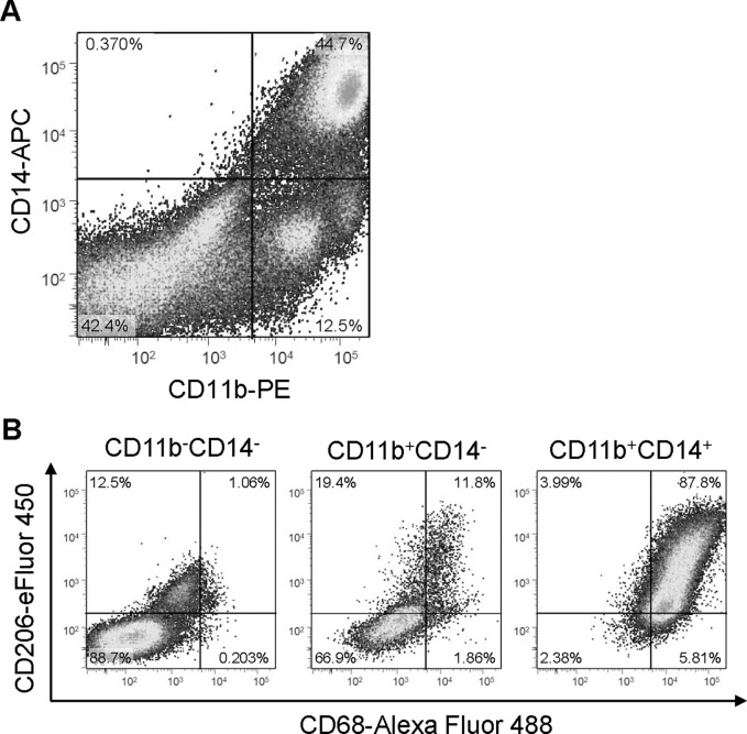
Fig 6. Characterization of primary cells from ovarian cancer ascites.
(A) FACS analyses. Ovarian cancer ascites were collected aseptically, and cells were isolated by standard Ficoll-Paque density-gradient. Thereafter, cells were labelled with cell surface markers CD11b (PE, x-axis) and CD14 (APC, y-axis). Cells were divided into 3 groups, CD11b-CD14-, CD11b+CD14- and CD11b+CD14+ cells. (B) The majority of CD11b+CD14+ cells are M2-polalized macrophages. These three populations of cells were further labeled with cell surface markers CD68 (Alexa Fluor-488, x-axis) and CD206 (eFluor-450, y-axis). 87.8% of CD11b+CD14+ cells (right) were CD68 and CD206 positive.

