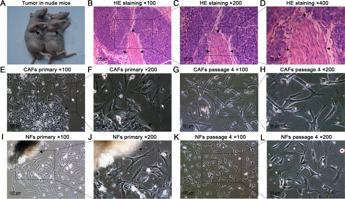
Fig 1. Tumor formation in nude mice, HE staining, and primary culture of CAFs and NFs.
(A) Four weeks after injection, tumor nodules formed in the right armpits of nude mice (black arrows). (B–D) HE staining confirmed that the nodules were typical laryngeal squamous cell carcinoma containing abundant CAFs (black arrows). (E,F) Two days after seeding, the CAFs (white arrow) and HEp-2 cells (black arrow) grew out of the tissue fragments. (G,H) At passage 4, purified CAFs were obtained. (I,J) The NFs grew out of the fragment. (K,L) At passage 4, purified NFs were obtained.

