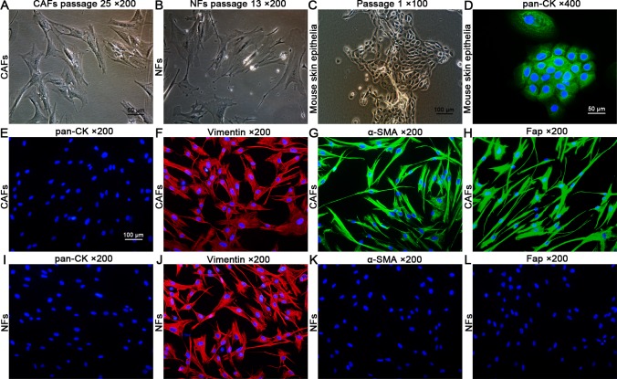
Fig 2. Cell morphology of CAFs, NFs, and mouse skin epithelia and immunocytochemical staining.
(A,B) Cell morphology of CAFs (passage 25) and NFs (passage 13). The CAFs did not show significant morphological changes compared with the NFs before passage 25; however, the NFs showed changes in morphology, including an enlarged and more flattened outline, compared with the CAFs after passage 10. (C) Cell morphology of primary cultured mouse skin epithelia at passage 1. (D) The mouse skin epithelia showed positive staining of pan-CK (positive control). (E–L) The CAFs and NFs showed negative staining of pan-CK and positive staining of vimentin, indicating their fibroblast identities. Compared with the NFs, the CAFs showed positive staining of α-SMA and Fap, two markers of activated fibroblasts.

