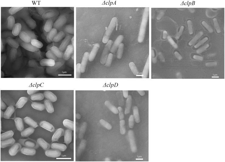
Fig 3. Scanning electron micrographs of wild-type and mutant biofilms.
Cells were grown for 24 h on LB plates and smeared on a silicon slice on the object stage. The samples were then imaged using a MIRA 3 scanning electron microscope at a magnification of 20000× times with a voltage of 15 KV. Scale bar = 1 μm.

