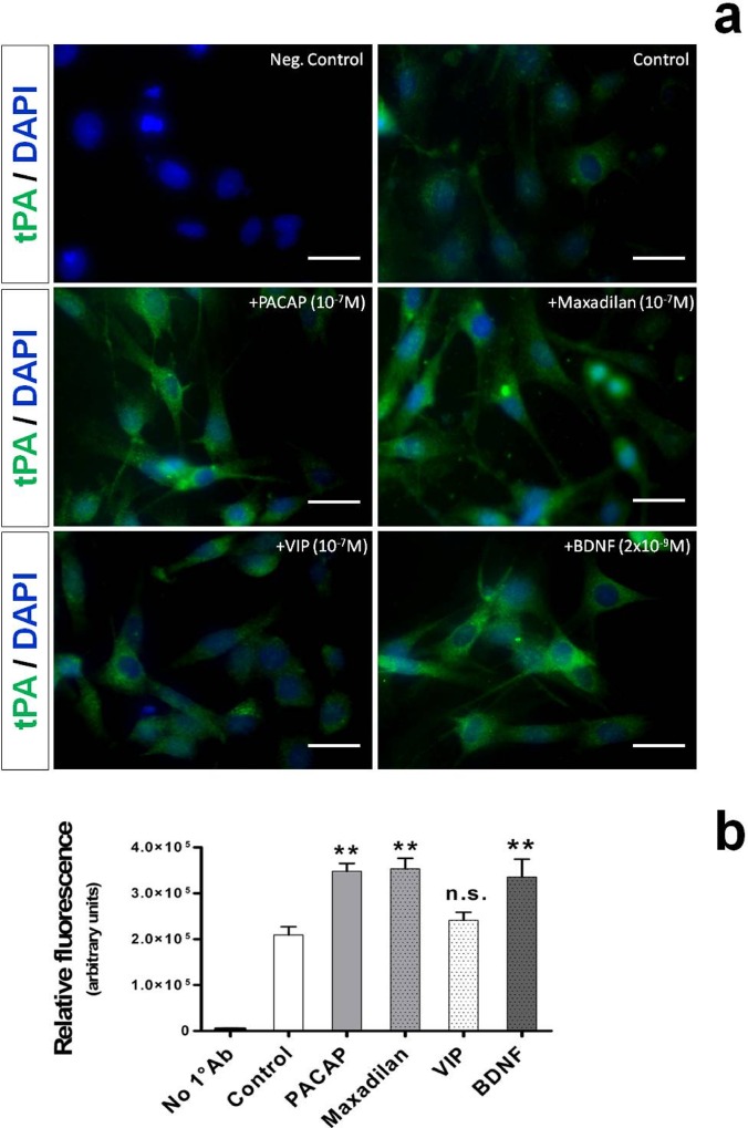
Fig. 2. Localization of tPA-like immunoreactivity in RT4-D6P2T cells stimulated with PACAP, maxadilan, VIP or brain-derived neutrophic factor (BDNF).
Depicted are representative immunocytofluorescence photomicrographs (a) and bar graphs showing semi-quantification of fluorescent intensity (b) in cells in which 1° Ab was omitted (Neg. Control), were left untreated (Control) or were treated with the indicated concentrations of PACAP, the PAC1 agonist (maxadilan), VIP or BDNF for 24h. Briefly, RT4-D6P2T cells cultured on glass cover slips were fixed in 4% para-formaldehyde, permeabilized with 0.2% Triton X-100, blocked with 0.1% BSA in PBS and then probed with the tPA antibody (1:50 dilution). Detection was then performed using an Alexa fluor 488-conjugated secondary antibody. Nuclei were counterstained with DAPI (#940110 Vector Laboratories). Immunofluorescent images were captured using an Axiovert 40 fluorescence microscope (Carl Zeiss Inc.). Each condition was reproduced in three dishes per experiment. Representative photomicrographs were taken from at least three fields per dish in a fixed pattern. Original magnification 40X. Scale bar = 30μm.

