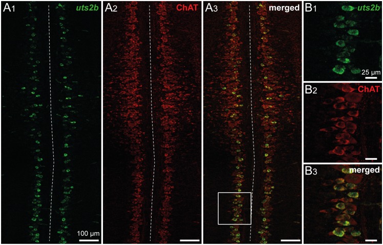
Fig 3. Spinal uts2b + cells are cholinergic neurons in X. laevis tadpoles.
Confocal images of combined fluorescent in situ hybridization of uts2b mRNA (A1, B1) and ChAT immunolabeling (A2, B2) in a whole-mount dissected and opened spinal cord preparation of stage 50 wild type tadpole. A3, B3. Merged image obtained when uts2b and ChAT stainings were superimposed. Dorsal view with rostral up. Note that in this preparation, the more ventral structures are close to the midline (dashed line). The boxed region in A3 is shown at higher magnification in B1–B3.

