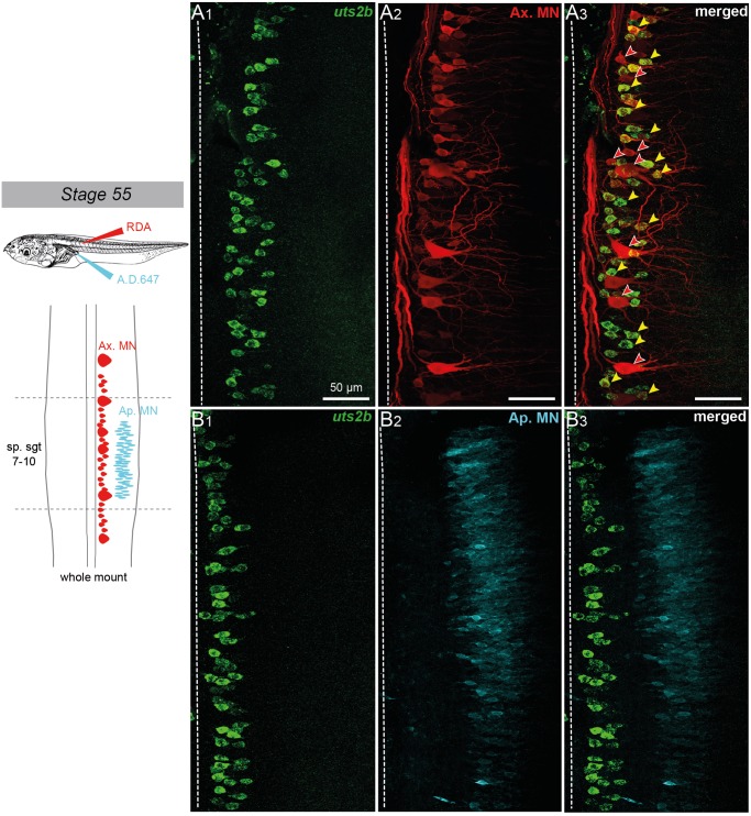
Fig 4. Spinal uts2b + cells of X. laevis tadpoles project to tail myotomes.
Confocal image of combined fluorescent in situ hybridization of uts2b mRNA (A1, B1) and retrograde labeling of spinal axial (A2) and appendicular (B2) motoneurons in stage 55 tadpole whole-mount spinal cord preparations. A3, B3. Merged images of uts2b staining and retrograde labeling. All images display dorsal view of hemi-cords, with the rostral side up (dashed-line on the left indicates the midline). Axial motoneurons (Ax MN) were labeled with rhodamine dextran dye (RDA) injected into tail myotomes while appendicular motoneurons (Ap MN) were labeled from posterior leg muscles with Alexa Dextran 647 dye (A.D. 647; see upper scheme on the left panel). The drawing on the left panel illustrates the localization of Ax MN and developing Ap MN in larval spinal cord at stage 55. Yellow arrowheads indicate double stained cells. Red arrowheads indicate uts2b—retrograde labeled cells. sp.sgt 7–10, spinal segments 7 to 10.

