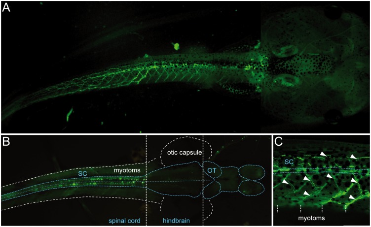
Fig 5. Most of the fluorescence visible in transgenic uts2b-GFP X. laevis tadpoles occurs in cells located in the spinal cord and in motor axon projections.
A. GFP fluorescence imaging of a representative transgenic tadpole at stage 58. The tail is seen in lateral view while the head is seen in dorsal view. B. GFP expression at the level of a dissected and opened whole-mount CNS of a stage 50 transgenic tadpole. The CNS was optically exposed by removing the dorsal part of the tail and the top of the head. Note that GFP+ cells are restricted to the spinal cord. Dorsal view, rostral to the right. C. Detail of A showing GFP+ motor axon projections (arrowheads) extending towards axial musculature. White dashed vertical lines indicate myotome boundaries. SC, spinal cord. OT, optic tectum.

