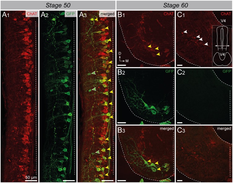
Fig 7. Spinal GFP+ cells of transgenic uts2b-GFP X. laevis tadpoles mainly express ChAT.
Confocal images of combined immuno-labeling of ChAT+ (A1) and GFP+ (A2) neurons in a whole-mount spinal cord preparation of stage 50 transgenic tadpole. A3. Merged image obtained when ChAT and GFP stainings were superimposed. Dorsal view with rostral up. Only the hemi-cord is shown. The dashed-line on the right side represents the midline. Note that in this preparation, the more ventral structures are closer to the midline. Confocal images of combined immuno-labeling of ChAT+ (B1–C1) and GFP+ (B2–C2) neurons in lumbar spinal cord (B) and brainstem (C) cross-sections of stage 60 uts2b-GFP tadpoles. B3, C3. Merged images obtained when ChAT and GFP stainings were superimposed. The cross-section in C1–3 originates from the caudal hindbrain (rhombomeres 7–8) where IX-XI motor nuclei are located. Yellow arrowheads designate GFP+/ChAT+ cells; green arrowheads designate GFP+/ChAT− cells; White arrowheads designate GFP−/ChAT+ cells. D, dorsal; M, medial; V4, 4th ventricule.

