
Fig 8. Spinal GFP+ axon motor projections of transgenic uts2b-GFP X. laevis tadpoles co-localize with α-bungarotoxin, a marker of postsynaptic neuromuscular junctions.
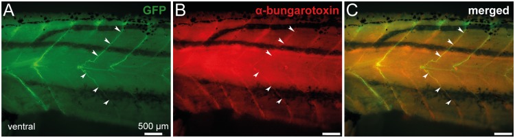
Combined fluorescence of GFP (A) and immuno-labeling of α-bungarotoxin (B) at the level of spinal axon motor projections in a whole-mount stage 57 transgenic tadpole. C. Merged image obtained when GFP fluorescence and α-bungarotoxin staining were superimposed. Postsynaptic nicotinic acetylcholine receptors labeled by α-bungarotoxin and GFP+ motor neuron axons overlap (arrowheads). Lateral views, rostral to the left.