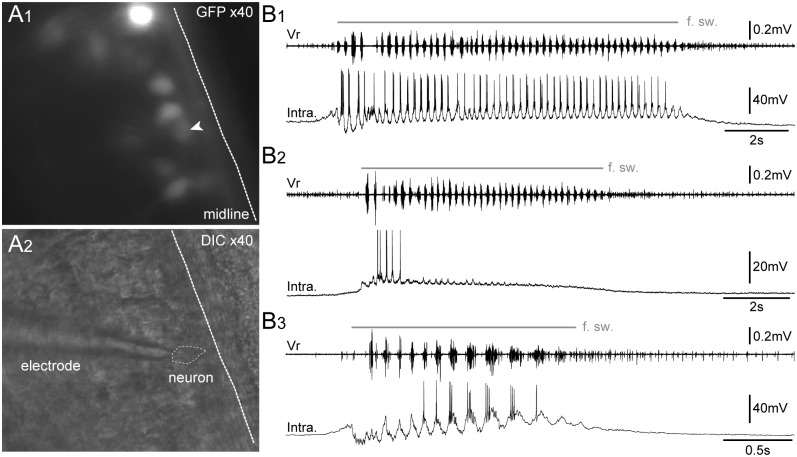
Fig 10. GFP+ cells exhibit a typical pattern of motoneuronal electrical activity during spontaneous fictive swimming episodes.
Visualization of spinal GFP+ neurons at x40 in fluorescence condition (A1) and infrared condition (A2) in a whole-mount isolated in vitro preparation of brainstem-spinal cord with dorsal-side opened. Patch-clamp intracellular recordings (bottom traces) of spinal GFP+ neurons (Intra.) either with a persisting (B1), a non-persisting firing pattern (B2), or a slow-delayed firing pattern (B3) during spontaneous fictive swimming episodes (f. sw.). The upper trace in B1–B3 represents the extracellular fictive swimming activity recorded simultaneously in a ventral motor root (Vr). DIC, differential interference contrast. The white arrowhead points to the GFP+ neuron targeted for patch clamp recording.

