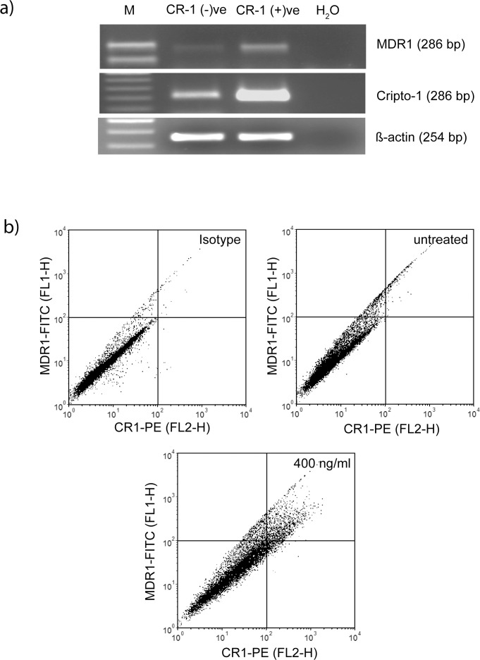
Fig 7. Co-induction of MDR-1 and CR-1.
a) U-87 MG cells treated with CR1-GST (400 ng/ml) for 24 hr were sorted in two subpopulations and RT-PCR was used to measure gene expression. b) Cells were treated with CR1-GST (400 ng/ml) or left untreated for 24 hr and expression of CR-1 and MDR1 was measured by flow cytometry. Cells in the right-upper quadrant are positive for both CR-1 and MDR1. Cells treated with CR1-GST (400 ng/ml) but stained with isotype control antibodies was used as negative control.

