
Fig 4. Rescue of TH+ and NeuN+ cells in the ipsilateral SN with intraperitoneal administration of anti-α-Syn antibodies.
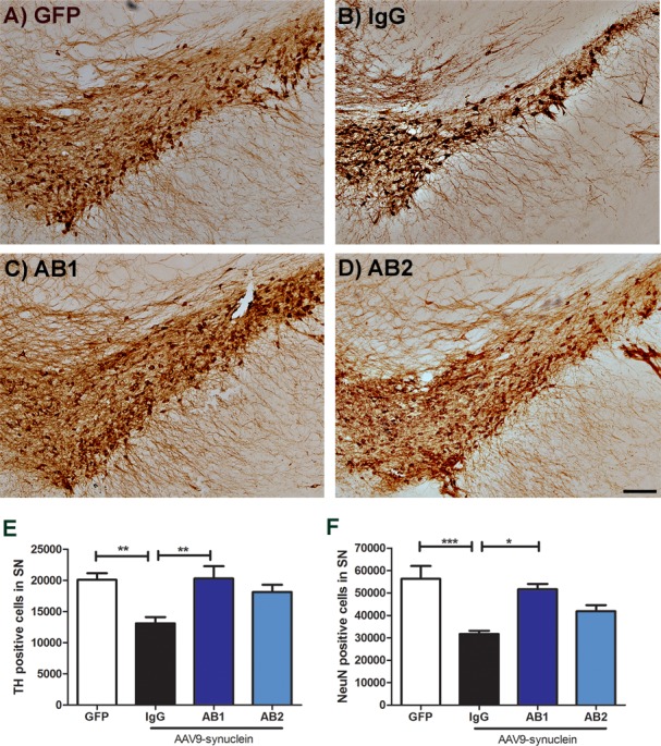
Immunohistochemical staining of the SN region with an anti-TH antibody (A) AAV-GFP, (B) AAV-α-Syn + IgG, (C) AAV-α-Syn + AB1, (D) AAV-α-Syn + AB2. (E) Graph of unbiased stereological estimation of TH+ cells in the SN of treated animals. AB1 treated animals showed similar levels of TH+ cells compared to the GFP control and significantly higher number of TH+ cells compared to the IgG treated group. Data are shown as mean ± SEM (n = 13 AAV-GFP control and n = 7 for treatment groups). (F) Graph of NeuN+ cells of the SN. Stereologic analysis shows a significant rescue of NeuN+ cells in SN sections with AB1 compared with IgG treated animals (n = 9 AAV-GFP control and n = 7 for treatment groups). *P< 0.05, **P< 0.01, ***P< 0.001. Scale bar = 100μm.