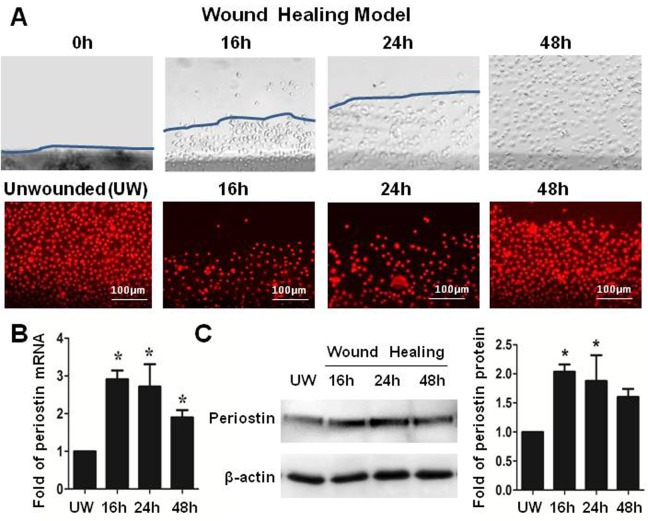
Figure 3. Periostin in an in vitro wound healing model of HLECs.
(A). Representative phase images immunofluorescent staining of PI (red) showing a 2-mm wide wound area was healed within 48 hours. (B). Reverse transcription-quantitative real-time polymerase chain reaction data showing the expression levels (relative fold of mRNA) of periostin by HLECs at different time points after wound. (C). Western blot results showing the protein levels of periostin by HCECs at different time points after wound. Data were shown as mean ± standard deviation, *p<.05; **p<.01, n = 3.

