
Fig 2. hMp84 stimulates cell death and stabilizes p53 protein in A375 melanoma cells.
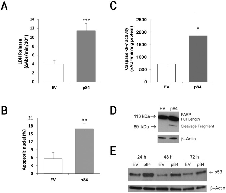
At 24 h after transfection, cell death was evaluated in EV-cells and in p84-cells as amount of intracellular LDH released in the culture medium (A), as percentage of nuclei showing an apoptotic morphology (Hoechst 33258 staining) (B), as caspase-3/-7 activation (enzymatic activity) (C), and as caspase-mediated proteolysis of poly(ADP-ribose) polymerase (PARP) (a typical Western blot experiment out of three is shown) (D). Results are presented as mean ± S.E. of three to six independent experiments. *P<0.05 and ***P<0.001, compared to control EV-cells. At 24, 48 and 72 h after transfection, p53 protein was evaluated by Western blot (E), in EV-cells and p84-cells. A typical Western blot out of three is shown.