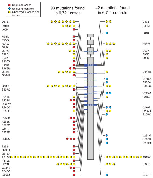
Figure 2. Apolipoprotein A-V (APOA5) mutations discovered after sequencing of 13,432 individuals.
Individual mutations (non-synonymous, indel frameshift and splice-site variants with minor allele frequency less than 1%) are depicted according to genomic position along the length of the APOA5 gene starting at the 5′ end (top). The number of circles on the left and right represents the number of times that mutation is observed in cases or controls, respectively. Dashed lines across the gene connect the same mutation seen in cases and controls. Mutations are shaded in red, blue, or yellow if observed in cases only, controls only, or both cases and controls, respectively.

