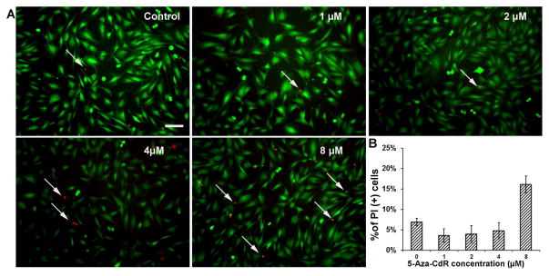
Fig. 1. Viability of MUCs following 5-aza-CdR treatment.
A. Calcein and PI were used to determine the viability of MUCs treated with control medium and medium containing 1–8 μM 5-aza-CdR. The viable and dead MUCs were stained with calcein (green) and PI (red) respectively and imaged using epifluorescence microscopy. Scale bar: 10 μM.
B. The percentage of viable cells in the control and 5-aza-CdR treatment groups. Approximately 93.08% ± 0.93%, 96.41% ± 1.62%, 96.06% ± 2.09%, 95.24% ± 1.97%, 83.89% ± 2.02% of MUCs were labeled by calcein when they were treated with medium containing vehicle, 1, 2, 4, and 8 μM 5-aza-CdR respectively. ANOVA suggested significant difference in the groups (P<0.05). Tukey post hoc test suggested that 8 μM 5-aza-CdR-treated MUCs showed a significantly decreased number of viable MUCs (P<0.05), while there is no significant difference among other groups.

