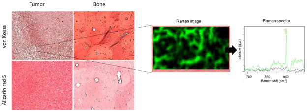
Figure 4.

Staining of MDA-MB-231 breast cancer samples and normal bone. With von Kossa (red/brown) and alizarin red S (red). The positive von Kossa stains indicate the presence of calcium carbonates suggesting the presence of Type II calcification specifically. A 100 × 60 μm2 region of the same tumor section scanned with a Raman microscope revelas the locations within that region where a 960 cm−1 shift was found (green color in the Raman image and Raman spectra) and where no shift near 960 cm−1 (black color). A shift at ~960 cm−1 indicates the presence of HAP.
