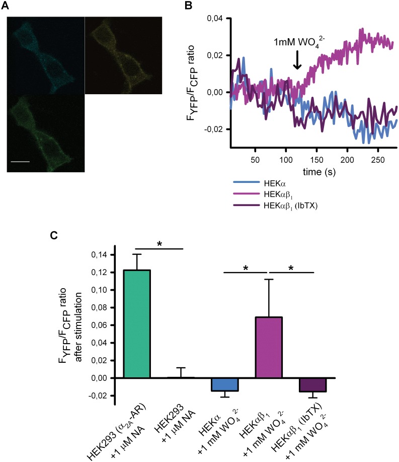
Fig 3. Tungstate-induced activation of heterologously expressed Gi/o proteins is mediated by BKαβ1 channels.
(A) Example of the reinforced membrane fluorescence pattern and emission levels from CFP channel (up-left), YFP (FRET channel) (up-right) and the merge channels (bottom-left) from HEK293 heterologously expressing Gαi-YFP and CFP-Gβ. Fluorescence microscopy images were recorded by using confocal microscopy (for more details see Methods). FRET signal was determined by using donor ratiometric parameters (458/514) after excitation in the CFP frequency and registering in the YFP emission frequency. (B) Representative FRET changes from HEK293, HEKα or HEKαβ1 cells transfected with the cDNAs of G protein subunits, in response to 1 mM tungstate (either in the absence or presence of IbTX), as indicated. (C), Average FRET changes for the different experimental conditions illustrated in B (n = 5–9). *P < 0.05 (Kruskal-Wallis test followed by Dunn post hoc test).

