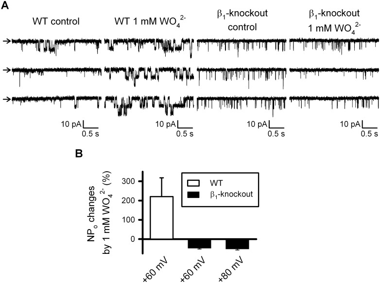
Fig 4. Tungstate-induced increase in the open probability of BK channels endogenously expressed in freshly isolated vascular myocytes requires the BK channel β1 subunit.
(A) Representative recordings obtained from inside-out patches clamped at + 60mV from wild-type (WT) and β1- knockout freshly isolated mouse vascular myocytes, before (control) and after exposure to 1 mM tungstate (1 mM WO4 2-), as stated. Arrows indicates the closed state level. (B) Average changes (in %) of BK channel open probability (NPo) induced by 1 mM tungstate on WT and β1- knockout mouse vascular myocytes at the indicated voltage membrane. Significant differences were found among WT (n = 7) and β1- knockout (n = 9), P < 0.001 (Student’s t-test). Further depolarization to +80 mV of β1- knockout inside-out patches did not change the response to tungstate found at +60 mV.

