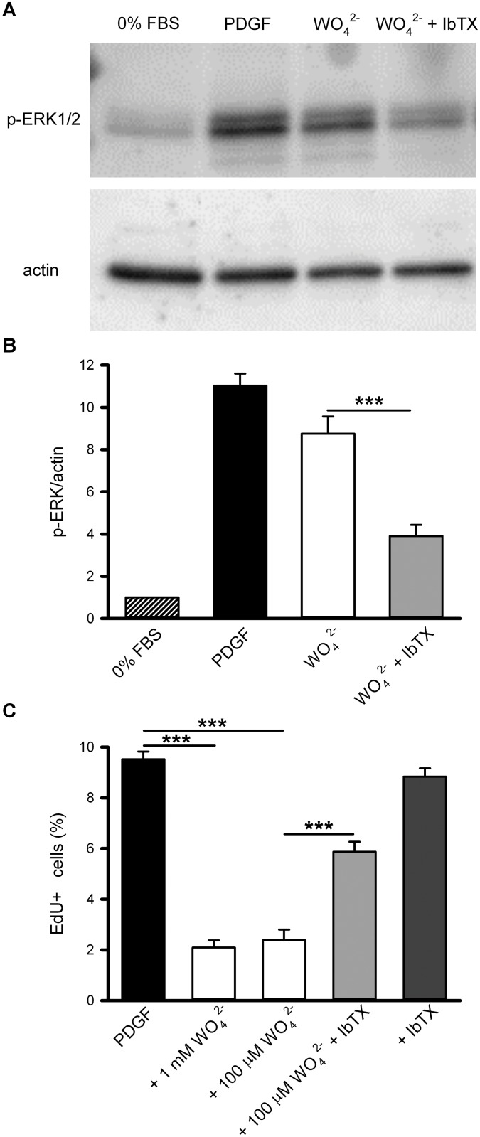
Fig 5. BK channels mediate both ERK1/2 phosphorylation and reduction of PDGF-stimulated proliferation induced by tungstate in human vascular smooth muscle cells.
(A) Representative Western-blot showing phosphorylation of ERK1/2 using phospho-ERK-specific antibodies after 10 minutes incubation in control conditions (0% FBS), with PDGF (20 ng/ml), and with tungstate (1 mM) alone or in combination with iberiotoxin (IbTX 100 nM), as indicated. Beta-actin was used as loading control. (B) Protein expression and phosphorylation were quantified by densitometry of the corresponding Western blot signal and normalized to beta-actin. The ratio p-ERK/beta-actin in control conditions was taken as 1, so that fold-changes relative to control are shown. (C) VSMCs were serum starved for 48 hours and then incubated 30 hours with 20 ng/ml PDGF alone or in combination with the indicated compounds. During the last 6 hours of incubation, EdU was added to the media to detect the number of cells entering S-phase. Mean ± S.E.M. of 6–9 determinations from at least four different cultures. ***P < 0.001 (ANOVA followed by Tukey post hoc test).

