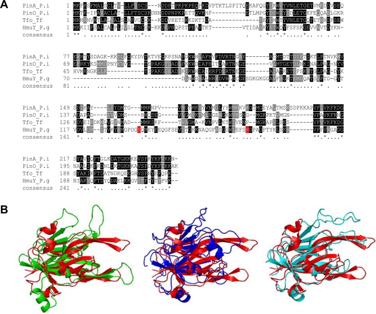
Fig 2. Comparison of P. gingivalis HmuY and its homologs from P. intermedia and T. forsythia.
Amino acid alignment (A) and approximated protein structures (B). P. intermedia PinA is shown in green, P. intermedia PinO in navy blue, T. forsythia Tfo in light blue, and P. gingivalis HmuY (PDB ID: 3H8T) in red. PinA, PinO and Tfo structures were modeled using Phyre2 modeling server and appropriate templates (PinA—PDB IDs: 3U22; PinO—PDB IDs: 3U22, 3H8T, 4GBS; Tfo—PDB IDs: 3U22, 3H8T, 4GBS).

