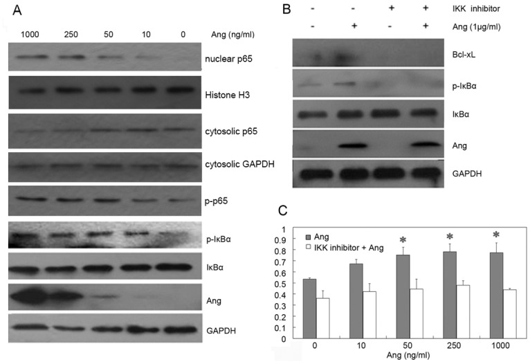
Fig 3. Activation of NF-κB pathway by Ang.

A. The cells were incubated with 0, 10, 50, 250, and 1000 ng/ml Ang for 2 h. The proteins of phosphorylated p65, phosphorylated IκBα, IκBα, Bcl-xL, Ang and GAPDH were detected in Ang-treated U87MG cells. The expression levels of p65 and Histone were detected in nuclear extraction preparations. The expression levels of p65 and GAPDH were detected in cytosolic extraction preparations. B. The cells were pretreated by the IKK inhibitor for 1h and were then stimulated by Ang at the final concentration 1μg/ml for 2h. After the protein was extracted, samples with equal amounts of protein were subject to SDS-PAGE and Western blotting analyses for phosphorylation of IκBα, IκBα, Bcl-xL, Ang and GAPDH. C. The inhibitor was added to 96-well plates containing 5,000 U87MG cells/well for 1 h before cells were stimulated by various concentrations of Ang. After a 48 h incubation, cell growth was measured.
