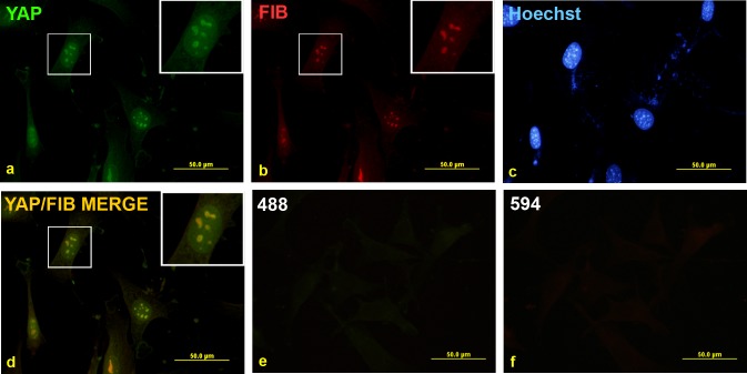
Figure 2. Punctate nuclear YAP staining is present in LPCs and co-localises with the nucleolar marker fibrillarin.
Non-transformed BMEL A-EGFP LPCs were grown on coverslips and fixed before being co-stained with YAP (a) and fibrillarin (FIB) (b) antibodies and counterstained with Hoechst stain as shown (c). Overlay of YAP and FIB images in (a) and (b) is shown in (d). Undetectable immunofluorescence was seen with the Alexa Fluor-488 (e) and-594 (f) secondary antibodies alone.

