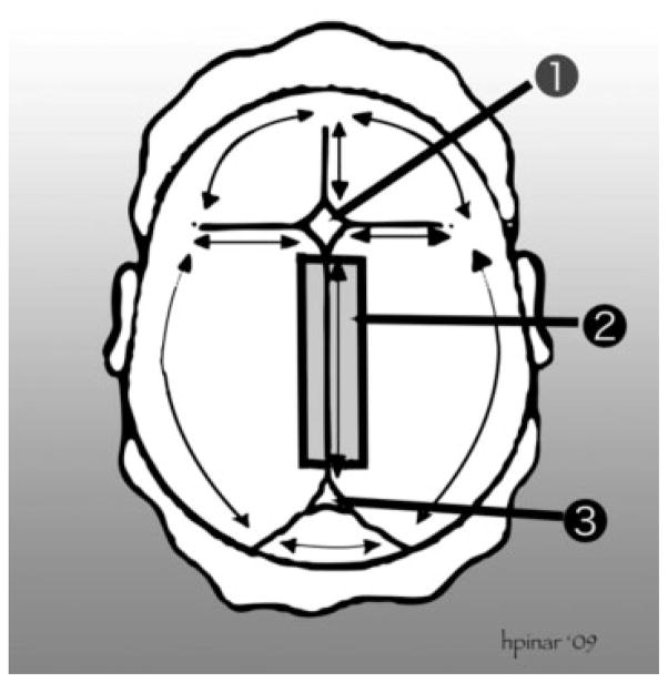
Figure 1.
Removal of the brain. This technique is suitable for fetuses of early gestations and involves cutting through the sutures and the connecting part of the bone flaps to remove the entire calvarium. (1) Anterior fontanelle. (2) The thick black lines represent the cutting lines to remove the sagittal sinus. (3) Posterior fontanelle. The scalp is incised starting behind one ear, extending to the back of the opposite ear. The first incision is made as far posterior as possible, in the area of the posterior fontanelle. The fetal scalp is soft and can easily be stretched without tearing. The scalp is then reflected downward to the level of the brows ventrally and the neck muscles dorsally. The cranial vault is opened by cutting through the membranous sutures. Cuts lateral to the sagittal suture are made to leave a strip of calvarial bone with the underlying sagittal sinus. Freeing the attachments of the brain is accomplished by turning the head on the occipital bone (or faceup) with the vertex directed to the prosector and both lateral flaps deflected. As the cranium is tilted gently to one side and then the other, all the cranial nerves and large vessels are identified and transected as close to the bone as possible, proceeding from front to back, and the anterior portion of the falx cerebri cut loose. As the posterior fossa is approached, the tentorium is cut away from the interior of the skull, around its entire perimeter, and then the falx cerebri is transected just above the tentorium. The cerebrum is then allowed to fall backward, and the pituitary stalk, anterior border of the tentorium, and the cervical spinal cord are cut as far distally as possible. With that, the entire brain slides out easily into a container and the specimen is weighed fresh. The external appearance of the brain is examined and any abnormalities are noted and photographed.28–30

