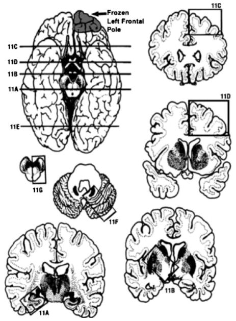
Figure 3.
Location of the brain slices and blocks obtained. Part of the left frontal pole was submitted fresh for research purposes. (11A) The first slice was at the level of hippocampus. (11B) Basal ganglia level. (11C) Frontal cortex and white matter. (11D) Parietal cortex and white matter. (11E) Occipital cortex and white matter. (11F) Cerebellum and pons. (11G) Medulla and midbrain. (11H) Cervical spinal cord.27–30

