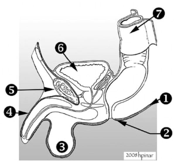
Figure 6.
Removal of the genitourinary system in toto. (1) Plane of dissection. This dissection is performed close to the skin so extra attention should be give not to damage the overlying skin. The plane of dissection is indicated by the dark thick line. (2) Anal opening. (3) Testes. (4) Penis. The skin over the penis is dissected and cut around the glans. (5) Symphysis pubis. (6) Bladder. (7) Rectum. In cases with suspected genitourinary abnormalities (cloacal dysgenesis, ambiguous genitalia, posterior urethral valves, anal atresia, or fistulas), dissection was performed at the caudal end of the organ block before it was separated from its pelvic attachments. After dissection of pelvic viscera, the pubic rami and the symphysis were then incised, so the ilia could be pushed laterally to open the bony pelvis. The entire length of the urethra and colon/rectum were thus freed from the pelvis and surrounding tissues. The organs connected to the base of the pelvic cavity were dissected as close as possible to the perineal skin. In males, the urethra was removed without disrupting the appearance of the external genitalia by using blunt dissection to free the cavernosa and glans penis from the penile skin. This skin segment was left intact and reexpanded with a small piece of gauze to return it to its normal outward appearance. The testes were removed with the organ block. Posteriorly, the rectum was dissected, as close to anal opening as possible, and resected with an ellipse of skin around it. This site should be sutured at the conclusion of the postmortem examination.

