
Figure 7.
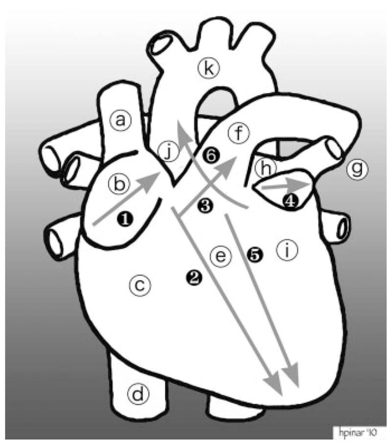
Dissection of the heart. Structures: (a) superior vena cava; (b) right atrial appendage; (c) right ventricle; (d) inferior vena cava; (e) septum; (f) pulmonary artery; (g) pulmonary veins; (h) left atrium; (i) left ventricle; (j) ascending aorta; (k) aortic arch. The cuts during dissection: (1) first cut opens the right atrial appendage; (2) second cut follows close to the interventricular septum all the way to the tip of the heart; (3) third cut goes through the pulmonary artery all the way to the splitting off the right and left arteries (pig’s snout appearance); (4) fourth cut opens the left ventricular appendage. After the initial small incision, this incision can be expanded so that the interior of the left atrium can be examined; (5) fifth cut opens the left ventricle adjacent to the interventricular septum; (6) sixth cut goes through the mitral valve, cuts the pulmonary artery above the valves, and enters into the ascending aorta.