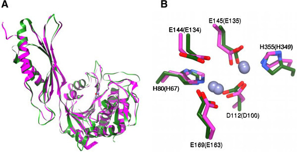
Figure 3.

Homology model of the [ZnZn(ArgE)] from E. coli based on the X-ray structure of DapE. A) Overlay of the ArgE homology model (Green) and the X-ray crystal structure of DapE (Magenta). The two Zn(II) ions and the bridging water from ArgE are shown as spheres. B) Conserved active site residues for ArgE (Green) and DapE (Magenta). Nitrogen atoms are in blue, oxygen atoms are in red. The residues are labeled with single-letter amino acid codes with the labels for residues from DapE in parenthesis.
