
Figure 2.
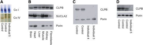
Analysis of CLPB Protein Levels and In-Gel Activity of Complexes I and IV
(A) In-gel enzyme activity in liver from individual 5 shows normal activity of complexes I and IV.
(B) Analysis by SDS-PAGE of CLPB protein in different human tissues showing ubiquitous localization of CLPB.
(C and D) Immunoblot analysis of CLPB in fibroblasts from individuals 3 and 4 and liver from subject 5 (D) shows absence of CLPB protein. In brief, mitochondrial protein was isolated from fibroblasts and liver as previously reported.23 The samples (25–30 cμg protein/lane) were run on a 12% SDS polyacrylamide gel and transferred to a PVDF membrane. The membrane was probed with a polyclonal antibody against CLPB (Atlas Antibodies) at a 1:1,000 dilution and developed with a 1:1,000 dilution of goat anti-rabbit antibody (Dako). An antibody against porin (Proteintech) was used as a loading control at a 1:1,000 dilution, and SUCLA2 was used as a reference at a 1:1,000 dilution. The secondary antibody was goat anti-mouse at a 1:1,000 dilution (Dako). The bands were visualized with the Supersignal West Pico and Femto substrates (Thermo Fisher Scientific) and MicroChemi imaging (DNR Bioimaging Systems).