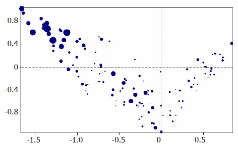
Figure 1.

MCA of exploratory set of 212 hepatocellular nodules in cirrhotic livers. The relative lesion size of the hepatocellular nodules is depicted as blue dots. The diameter of the dots varies according to the size of the nodule observed. Size does not seem to allow a classification of nodules, in other words, HCC can be of all sizes, and large nodules are usually HCC. However, small nodules are not necessarily MRN or DN.
