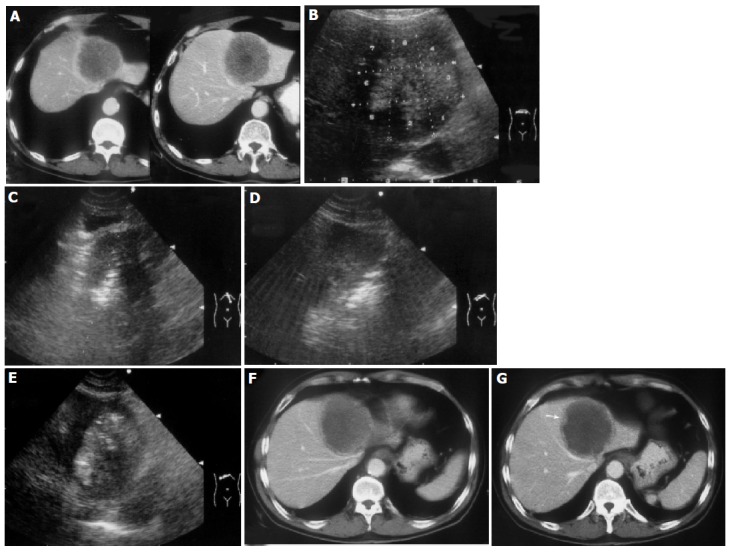
Figure 1.

A 55-year-old man developed liver metastasis from colon cancer. A: Enhanced transverse CT scans obtained before treatment showed a spherical tumor invading into the diaphragm; B: The ablation protocol was designed and eight target sites were determined in this section. The tumor area near the heart and stomach were ablated first. C: The fourth ablation was conducted; D: The fifth ablation was conducted; E: The ablation was finished and the tumor area was presented as hyperechoic; F: Enhanced transverse CT scans obtained 1 d after the treatment showed that most of the tumor was coagulated and necrosis; G: Enhanced transverse CT scans obtained 1 d after the treatment showed slight enhancement (↑) on the lower margin of the tumor. The patient received the second treatment and achieved complete ablation.
