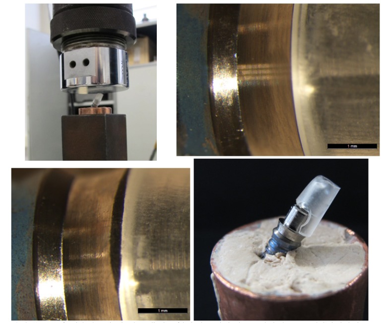
Figure 2.
A) Image of static load test showing the application of the load onto the implant-prosthetic abutment. B&C) Optical microscope images of condition of a Group MP abutment pre- and post-loading. No differences were registered at the implant-abutment union. D) Image of Group MP abutment showing the methacrylate fracture at the interface with the machined titanium base.

