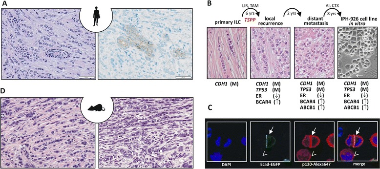
Figure 1.

Infiltrating lobular breast cancer, a human infiltrating lobular breast cancer cell line and a genetically engineered mouse model for infiltrating lobular breast cancer. (A) Representative photomicrographs of infiltrating lobular breast cancer (ILC) stained with hematoxylin and eosin (left) or subjected to immunohistochemistry for E-cadherin (right). Note the E-cadherin-positive normal mammary gland duct surrounded by E-cadherin-negative ILC cells. (B) Molecular evolution of the IPH-926 ILC cell line. Photomicrographs show the histomorphology of the corresponding clinical tumor specimens and the IPH-926 cell line in vitro. Arrow highlights a single file linear cord reminiscent of primary ILC. AI, aromatase inhibitor; CTX, various conventional chemotherapies; LIR, local irradiation; TAM, tamoxifen; TSPP, transition to a secondary pleomorphic phenotype; yrs, years; M, mutational inactivation; ↑, overexpression; ↓, loss of expression. (C) Reconstitution of E-cadherin in ILC cells induces relocation of p120-catenin (p120) to the cell membrane. Shown are fluorescence images of IPH-926 cells transiently transfected with an E-cadherin–enhanced green fluorescent protein (Ecad-EGFP) expression construct and stained with an anti-p120-Alexa647 antibody. Closed arrow, cells with ectopic expression of Ecad-EGFP; open arrow, a cell without Ecad-EGFP. Note the accentuated membranous p120 staining in cells expressing Ecad-EGFP. DAPI, 4′,6-diamidino-2-phenylindole. (D) Mouse ILC from genetically engineered mouse models. Left, a tumor reminiscent of classic ILC; right, a pleomorphic mouse ILC. Both micrographs generated from the WAPcre;Cdh1 F/F ;Trp53 F/F mouse ILC model.
