Figure 4.

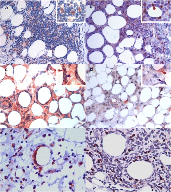
Immunohistological confirmation of the protein expression of the up regulated genes in SPTL. a) CXCL9-expressing, morphologically mostly malignant lymphocytes in a SPTL lesion (red, 20x). b) IDO-1-expressing morphologically malignant lymphocytes (red arrow) rimming a fat cell in a SPTL lesion (red, 20x). c) Double immunostaining for CD8 (cyan) and CXCR3 (red) showing CD8+CXCR3+ lymphocytes (red arrow) in a SPTL lesion (20x). Cells expressing only CD8 are indicated with blue arrow. No counterstain. d) Double immunostaining for CD8 (cyan) and CXCR3 (red) showing exclusively the expression of CD8 and CXCR3 in different cells in a LEP lesion (20x). No counterstain. a)-d) Insert in upper right corner represents magnification of 40x. e) CXCR3-expressing malignant lymphocytes rimming the fat cell in a SPTL lesion (red, 20x). f) High number of FoxP3+ (brown) regulatory T-cells in a SPTL lesion (red, 40x).
