
Figure 3.
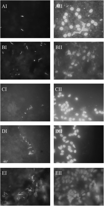
Adhesion of LAB20 to the canine IECs. Adherence of FITC-labelled bacteria to canine IECs obtained from duodenum (A), jejunum (B), ileum (C), cecum (D), and colon (E) sections is shown in the left panel. The arrows indicate LAB20 cells adhered to IECs. The nucleus of IECs were stained with DAPI and are shown in the right column.