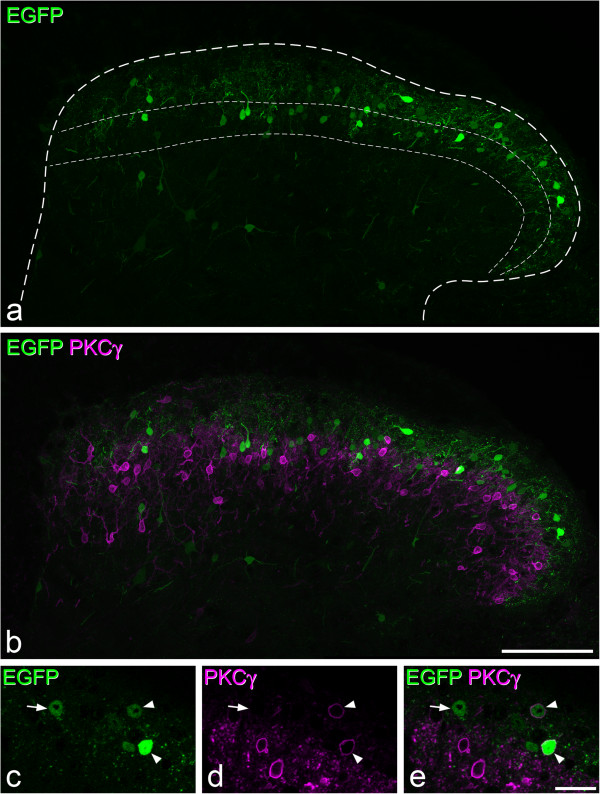
Figure 6.

Distribution of EGFP cells in the GRP-EGFP mouse. a, Immunostaining for EGFP (green) in a transverse section shows scattered cell bodies throughout laminae I-II (projection of 20 optical sections at 1 μm z-spacing). The outer dashed line represents the outline of the dorsal horn, while the region between inner two lines corresponds to the inner half of lamina II, as defined by the plexus of PKCγ+ dendrites. b shows the same field scanned to reveal EGFP and PKCγ (magenta). Note that these are expressed in largely non-overlapping neuronal populations. c-e, Immunostaining with antibodies against EGFP (green) and PKCγ (magenta) showing the inner part of lamina II in a single optical section. Two EGFP+ cells that show weak PKCγ-immunoreactivity are indicated with arrowheads, and an EGFP+ cell that lacks PKCγ is marked with an arrow. Scale bars = 100 μm (a,b) and 20 μm (c-e).
