
Figure 2.
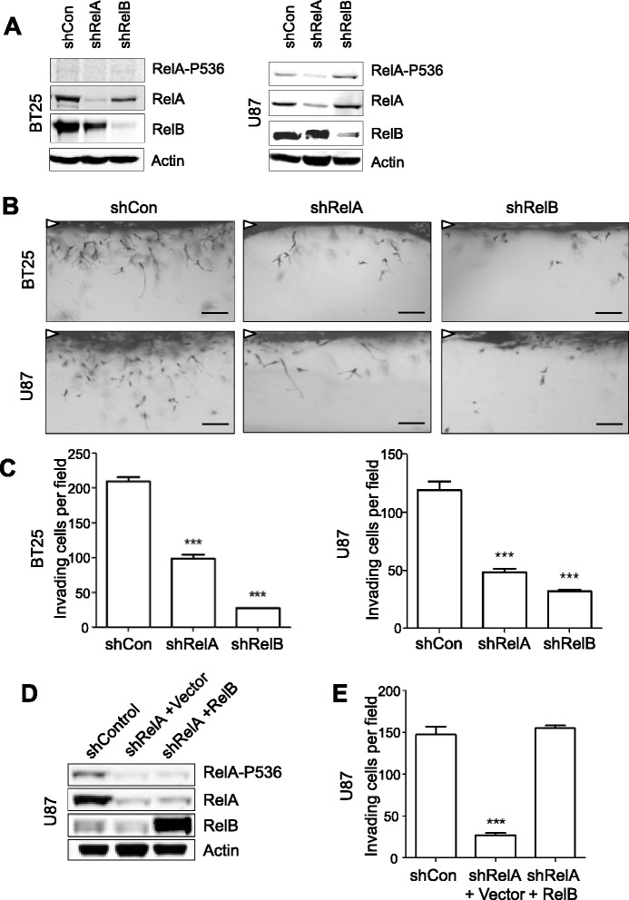
RelB poteniates glioma invasion independently of RelA. (A) Western blot of whole-cell lysates from NSC-cultured BT25 and U87 cells transduced with lentiviral vectors expressing shRNA targeting RelA (shRelA), RelB (shRelB), or a scrambled control (shCon). (B) Representative images of invading shRNA-transduced glioma cells at 48 h. White arrowheads indicate monolayer. Scale bars = 100 μm. (C) Quantification of invasion density at 48 h for BT25 and U87 knockdown cell lines. Data represent average numbers of invading cells per 1-mm2 field (n = 3 wells) ± S.E.M. ***p < 0.001 relative to shCon using One-way ANOVA with Tukey’s H.S.D. post-test. (D) Western blot of whole-cell lysates from U87 shRelA cells transduced with lentiviral vectors containing RFP cDNA (+Vector) or RelB cDNA (+RelB). (E) Quantification of invasion density at 48 h for U87 shRelA cells in Figure 2D. ***p < 0.001 relative to shCon using One-way ANOVA with Tukey’s H.S.D. post-test.