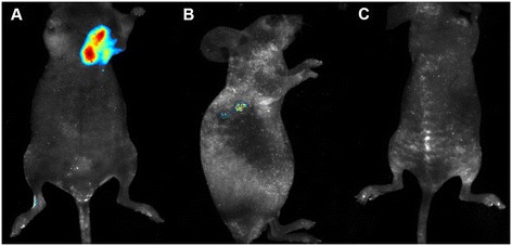
Figure 8.

Representative xenograft tumor nude mice models of Cervical carcinoma in vivo imaging. (A) In vivo fluorescence imaging 24 h after a subcutaneous injection of 5 mmol/L Au/Ce NCs solution near the tumor. (B) In vivo fluorescence imaging 24 h after a intravenous injection 5 mmol/L Au/Ce NCs solution through the tail. (C) Control nude mice without tumor after a intravenous injection equivalent PBS through the tail. Fluorescent Au/Ce NCs were observed inside the tumors using a 455 nm excitation wavelength.
