
Figure 1.
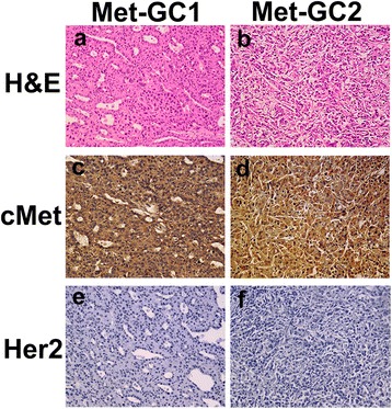
Histological and molecular characterization of the PDTX models of gastric cancer. a, b: H&E staining. c, d: Immunohistochemical staining for cMet. e, f: Immunohistochemical staining for Her2. Immunodetectable protein is indicated by brown staining; nuclei are counterstained blue. Original magnification, ×200.