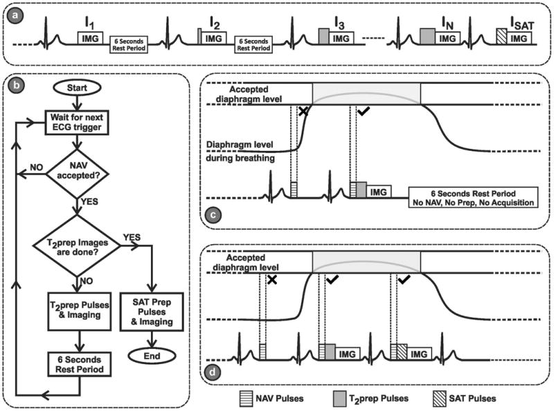
Figure 1.

(a) The schematic of the proposed sequence. Multiple single-shot images of the heart are acquired using ECG-triggering, following T2prep of different echo lengths, TET2P. Between each image, a 6 second rest period (with no RF pulses) is applied to allow for full re-growth of the myocardium signal. An image, ISAT, is acquired directly after a saturation pulse to simulate the effect of a very long T2prep echo time (i.e. T2prep = ∞) for improved estimation of the third parameter in the 3-parameter fit. (b) Flowchart for the proposed navigator (NAV)-gated acquisition scheme, The NAV is placed before the T2prep. If the NAV signal preceding the acquisition of the kth image is outside the gating window, no T2prep or imaging pulses are applied, leaving the magnetization undisturbed, and the acquisition of this image is repeated in the next R-R interval. If the NAV signal is within the gating window, the image with the desired T2prep time is acquired, followed by a 6 second rest period for magnetization recovery. (c) An example of the rejection-reacquisition scheme for a T2-prepared image. (d) An example of the acquisition of a saturation-prepared image, which immediately follows the T2-prepared image without any rest periods, and where the NAV is placed before the saturation pulse.
