
Figure 5.
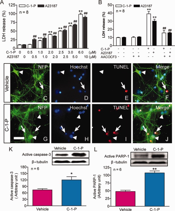
Effects of cytosolic phospholipase A2 (cPLA2) activation on spinal cord neuronal death in vitro. (A) Cultured spinal cord neurons were treated with the designated concentrations of ceramide-1-phosphate (C-1-P) or A23187 for 24 hours. Lactate dehydrogenase (LDH) release assay revealed that both cPLA2 activators, C-1-P and A23187, induced cultured spinal cord neuronal death in a dose-dependent manner. **p < 0.01, ##p < 0.01 versus the vehicle control, 1-way analysis of variance (ANOVA), Tukey post hoc test; n = 8. Data are shown as the mean ± standard error of the mean (SEM) from 3 independent experiments. (B) Importantly, neuronal death induced by C-1-P (2μM) or A23187 (5μM) was significantly reversed by AACOCF3 (15μM), a cPLA2 inhibitor. AACOCF3 was added 30 minutes before C-1-P or A23187, and the culture medium of each well was removed at 24 hours after the activator treatment for LDH release assay. **p < 0.01 versus the vehicle control, ##p < 0.01 versus the C-1-P or A23187 group, 2-way ANOVA, Tukey post hoc test, n = 8. Data are shown as the mean ± SEM from 3 independent experiments. (C–J) Administration of C-1-P (2.5μM) for 2.5 hours in vitro induced massive neuronal apoptosis (G–J) as compared to the vehicle controls (C–F). Apoptotic cells were identified by both Hoechst and triphosphate nick-end labeling (TUNEL) staining (arrows) and normal neurons were identified by staining for neurofilament protein (NFP) and Hoechst (arrowheads). Bar = 30μm. (K, L) C-1-P (2.5μM) for 2.5 hours also induced significant increase in the expression of active caspase-3 (K) and poly(adenosine diphosphate ribose) polymerase 1 (PARP-1) (L), two important components in the apoptotic pathway. *p < 0.05, **p < 0.01 versus the vehicle control (Student t test); n = 6. Data are shown as the mean ± SEM from 3 independent experiments.