
Figure 4.
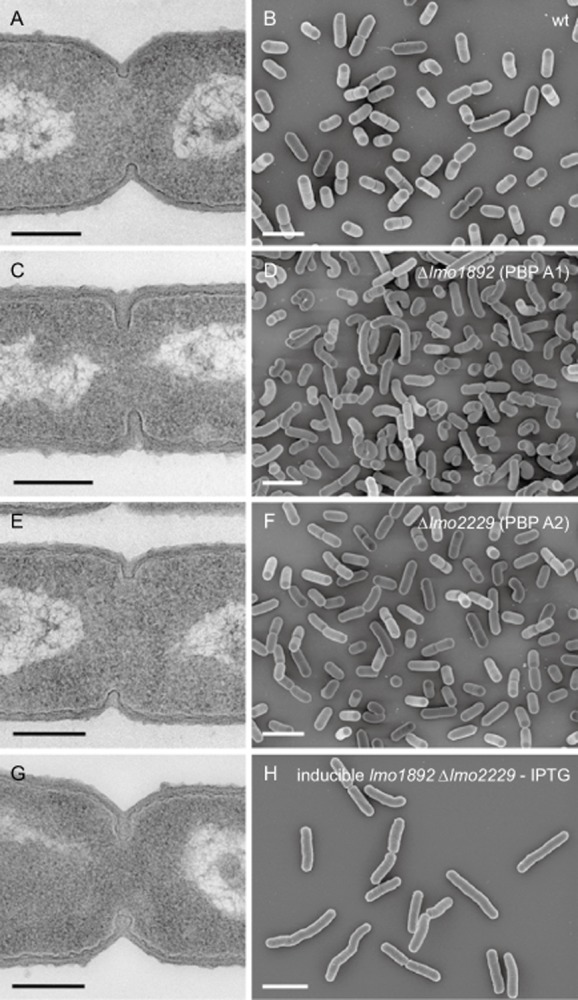
Effect of PBP A1 and PBP A2 inactivation on cell morphology. Transmission electron microscopy of ultrathin sections (A, C, E, G) and scanning electron microscopy (B, D, F, H) of fixed whole cells of L. monocytogenes strains devoid of class A high molecular weight penicillin-binding proteins. L. monocytogenes strains EGD-e (wt), LMS57 (Δlmo1892), LMS64 (Δlmo2229) and the inducible double mutant strain LMJR30 (Ilmo1892 Δlmo2229) were grown to mid-logarithmic growth phase in BHI at 37°C and subjected to chemical fixation and subsequent electron microscopy as described in the experimental procedures section. Scale bars: left column 200 nm, right column 2 μm.