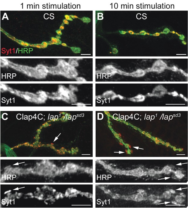
Figure 4. Acute inactivation of LAP results in the appearance of abnormal membrane structures that contain SV proteins.

Third instar larval NMJ preparations from the indicated genotypes were dissected, incubated with FlAsH-EDT2 and muscle 4 from segment A2 or A3 was exposed to 488-nm-filtered fluorescent light for 5 min. Following illumination, the larval NMJs were stimulated with high K+ (90 mm) Jan's solution for either 1 min (left column) or 10 min (right column). Following nerve stimulation, the larval body wall NMJ preparation was washed rapidly with Ca2+-free HL-3 saline and fixed. The larval NMJs were then stained with antibodies to HRP (neuronal membrane) and to the SV protein, Syt1. In control flies (A and B), Syt1 is found to be localized within the boutons in a typical ‘doughnut’ pattern. However, when LAP is inactivated (C and D), Syt1 distribution becomes more punctate. These punctate are sometimes localized to HRP-positive areas that appear to protrude outward from the boutons (arrows). Scale bars = 10 µm.
