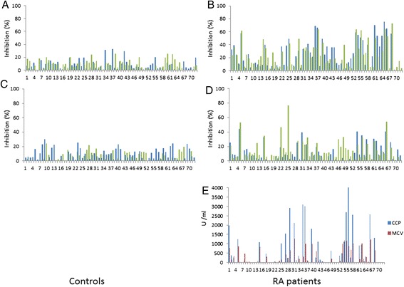
Figure 2.

Overview of the antibody specifities in controls (panels A and C) and RA patients (panels B and D) and RA patients’ CCP and MCV results. In panels A and B, antibodies specific to CitI are depicted in dark columns/blue and those specific for HcitI in light columns/green. In panels C and D, antibodies to CitII are presented in dark columns/blue and those to HcitII in light column/green. The values shown are percent of inhibition by respective soluble peptides to binding to the similar immobilized peptide. Panel E shows the binding of RA patient sera to the antigens of the CCP and MCV assays, shown as arbitrary units defined by each manufacturer. CCP, cyclic citrullinated protein; Cit, citrulline; Hcit, homocitrulline; MCV, mutated citrullinated vimentin; RA, rheumatoid arthritis.
