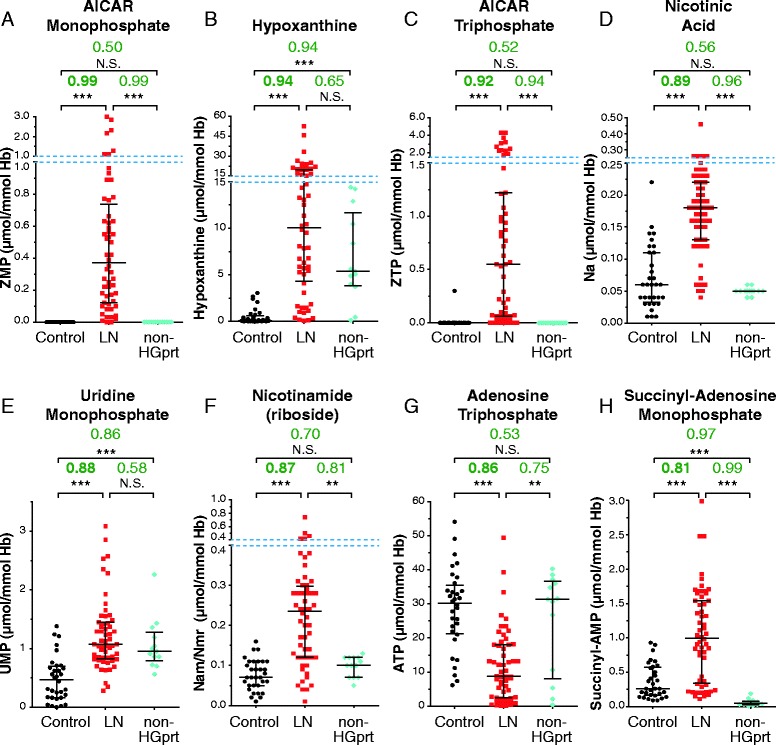
Figure 4.

Identification of the metabolites significantly changed in red cells of HGprt deficient patients. (A-H) For all categories, each dot corresponds to the mean of metabolite content measured in independent red blood cell extracts. AUC values (green numbers) correspond to Area Under Curves values deduced from ROC (Receiver Operating Curves) analyses (Additional file 1: Figure S1) performed as described in Methods. p-values were obtained from a Mann–Whitney–Wilcoxon test. NS: non-statistically different = p-value > 10−1; *: p-value < 10−2; **: p-value < 10−3 and ***: p-value < 10−4. Control: healthy patients (black circles); LN: Lesch-Nyhan patients (HRH + HND + LND; Red squares); Non-HGprt: non HGprt-deficient patients with hyperuricemia (blue diamonds). Nicotinamide (riboside) stand for the mix of nicotinamide and nicotinamide riboside, these two metabolites being not separated under our chromatographic conditions. Raw data are presented in Additional file 1: Table S1. Dashed blue lines indicate scale breaks.
