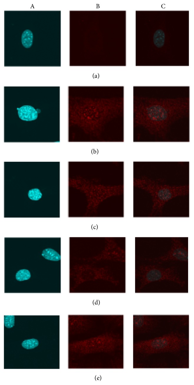
Figure 7.

The effect of ICA on NF-κB (p65) nuclear translocation was qualitatively assessed on young mouse aortic ECs. Magnification ×650 times. (a) shows the nuclei in aortic ECs by DAPI stain. (b) Red fluorescence signals (represent NF-κB p65 protein) were shown in aortic ECs both including the areas of nuclei and cytoplasm. (c) The combination of figure A and figure B helps to determine the red fluorescence signal in the nuclei and thereby indirectly showing NF-κB (p65) nuclear translocation from cytoplasm to nuclear. (a) Young aortic EC group (PBS and DMSO treatment). (b) Young + TNF-α group. (c) Young + TNF-α + ICA. (d) Young + TNF-α + ICA + PDTC. (e) Young + TNF-α + ICA + PDTC + Nicotinamide. As shown in the Figure 7(a), in the quiescent condition treatment with PBS or DMSO, very low red fluorescence signals were detected in aortic ECs and in the area of nuclei almost no red fluorescence was detected. After being treated by TNF-α to induce cell inflammation, red fluorescence signals were strongly detected in the cell, especially condensed in the nuclear location Figure 7(b). In the presence of ICA or ICA + PDTC intervention, such condensed red fluorescence signals in the nuclei were both reduced and ICA + PDTC intervention showed a stronger inhibitory effect than ICA, in Figures 7(c) and 7(d), respectively. As shown in Figure 7(e), adding Nicotinamide (SIRT6 enzyme inhibitor) red fluorescence signal became stronger than that in Figure 7(d).
