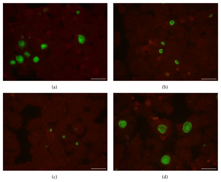
Figure 4.
Immunohistological staining of C. trachomatis infected cell monolayers in the presence or absence of EOMS. HeLa cell monolayers were infected with C. trachomatis (MOI 0.05) and incubated in the presence ((a) 16 μg/mL; (b) 32 μg/mL; and (c) 64 μg/mL) or absence of EOMS (d). After 48 h of incubation, HeLa cell monolayers were fixed, stained, and visualised by fluorescence microscopy (400x magnification). Bars: 50 μm.

