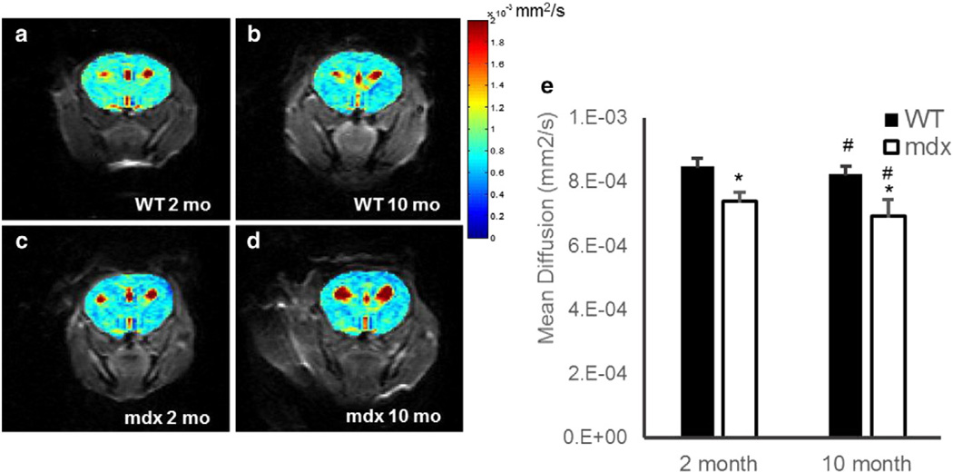
Fig. 1.
a–d. Representative diffusion images of 2-month-old WT (a), 2-month-old mdx (b), 10-month-old WT (c), and 10-month-old mdx (d). e. Mean cerebral (excluding ventricles) diffusion in C57/BL6 WT (black) and mdx (white), respectively. There was a significant decrease in mean diffusion as compared to WT observed in the mdx in both the 2- and 10-month-old mice (*p < 0.0005). There was also a significant age-dependent decrease in diffusion in young versus aged WT and mdx mice, respectively (#p < 0.05). Data represented as mean ± SD. Color bar is uniform in each panel.

