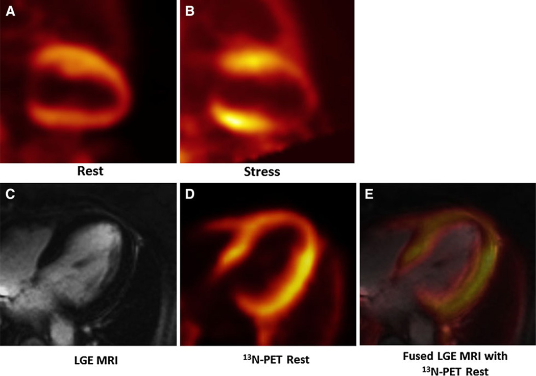
Figure 2.
13N-ammonia and late gadolinium enhancement (LGE) PET/MR. A, B Two-chamber long axis views of rest (A) and stress (B) 13N-ammonia PET/MR MPI demonstrating a predominantly reversible anterior/anteroapical perfusion defect consistent with ischemia. C LGE imaging shows no delayed contrast uptake in the anterior/anteroapical wall, indicating the absence of infarction. D 13N-ammonia PET demonstrating mild anterior and apical hypoperfusion at rest. E Fused PET perfusion and MR LGE images show no LGE in the area of resting hypoperfusion detected by PET.

